Research: Current Projects
Autism across the lifespan
Imaging studies in toddlers: Finding early brain markers
Dr. Inna Fishman, who directs the SCANgroup at BDIL, and her colleagues at SDSU, UCSD, and Rady Children’s Hospital (Drs. Ralph-Axel Müller, Ruth Carper, Themba Carr) are conducting a research study to identify early brain markers for autism spectrum disorders. This project is funded by the National Institutes of Mental Health and aims at detecting unique “brain signatures” in the first years of life in toddlers with ASD compared to typically developing children.
As a disorder of brain development, autism affects how the brain grows and works; yet, brain development trajectories accompanying the emergence of first symptoms of ASD, in the 2nd year of life, are almost completely unknown. This research study aims to identify how brain networks are organized and change over time, during the most critical developmental window when ASD symptoms first emerge (at age 15-26 months) and reach their peak (at 4-5 years of age). Identification of brain markers of risk will advance future discoveries of novel, effective diagnostics and treatments in ASD. Experts agree that better and earlier detection of autism is critical for successful treatment.
Another recently begun imaging project similarly studies children with ASD from age c.18 months into the preschool years, led by Drs. Annika Linke and Ralph-Axel Müller. This study additionally focuses on the development of the auditory system and sound processing. It aims to answer the question whether early auditory processing is atypical in toddlers with ASD and how this may affect language development.
If you have a 15-36 months old child with a diagnosis of ASD (or suspect she or he may have one), or if you have a typically developing toddler, please consider taking part in these important projects. If you are unsure about your child’s status, our experienced developmental psychologist will conduct a play-based diagnostic evaluation to assess whether your child may have ASD. Should your child be eligible for the study, you will be asked to bring your child for an MRI scan around his or her bedtime. Your child will be scanned while naturally asleep (no sedation will be involved). To achieve the goals of the study, we would also like to see you again up to 2 more times before your child turns 5 years old (each time will be about 1.5 years apart). You will be compensated for your time (you will earn $100 for the MRI visit and $50 for the evaluation / introductory visit).
Click here to find out more.
Imaging in preschoolers and young children: The “Toy Study”
Atypical responses to sensory stimuli like touch are common in autism. While ASD is comprised of both sensory and social deficits, the impact of atypical haptic (i.e., touch) and multisensory (e.g., vision and touch combined) processing on social and communicative abilities remains unknown; it is not well-understood how sensory processing may predict later social impairment in children with ASD. This NIMH-funded study, led by Dr. R. Joanne Jao Keehn, investigates the development of vision and touch in young children with ASD compared to typically developing peers at 4-5 years and 6-7 years of age, and how these sensory modalities can affect social outcomes in children with ASD. The findings from this research study will enhance our understanding of the biological bases of ASD and identify sensory abnormalities associated with sociocommunicative abilities, particularly in young children with ASD for whom early interventions can mitigate symptom severity.
Our interactive study involves looking at and feeling 3D printed objects and toys. We are using a longitudinal and multimodal approach that combines several state-of-the-art methodologies, including behavioral task measures, functional magnetic resonance imaging (fMRI), resting state functional connectivity MRI (fcMRI), diffusion weighted imaging (DWI), and neuropsychological assessments of social, sensory, and cognitive functioning.
We are seeking both typically developing children and children with a diagnosis of ASD aged 4 to 7 years old to take part in this exciting project. If eligible, your child’s participation will involve 3 visits: 1) a play-based developmental evaluation, 2) looking at and feeling objects and toys, and 3) a brain scan. Depending on your child’s age, we may ask you to repeat these visits once more in about 1 year. Participants will receive $125 for completing all testing (approximately 5 hours total across 3 visits), as well as a small toy.
Imaging in school-age children and adolescents

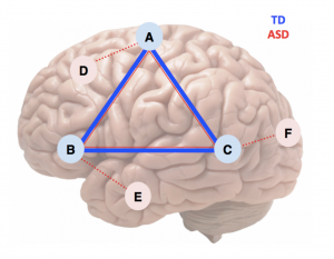
Simplified illustration of general pattern in ASD. Connectivity between 3 regions of a specialized network (A-B-C) is less strong in ASD than in typically developing (TD) children, indicated by the relatively thin red line. However, the network shows additional connections with other parts of the brain (D, E, F) in ASD (dotted red lines), whereas in the TD brain these connections have been “pruned” during development.
The findings are too numerous to describe one by one, but one main conclusion should be mentioned: The popular notion that in ASD the brain is “underconnected” is misleading. Our group has, for more than a decade, published studies showing that – on the contrary – brain regions are often overconnected (“talk too much to each other”). The exact pattern of findings is complicated (and in some cases differs from study to study).
However, a general pattern emerges. Functional brain networks include regions that are involved in a specific kind of task. For example, we have a language network that matures in childhood and includes regions in different parts of the brain (such as Broca’s area in the frontal lobe and Wernicke’s area in the temporal lobe). In neurotypical development, such regions that share a process become more strongly connected; but at the same time, they may lose connections (through so-called “synaptic pruning”) with other regions and networks that are involved in unrelated functions (e.g., vision). Networks therefore become more distinctly “sculpted” throughout development. This process seems to be affected in ASD. Connections within specialized networks are weaker (underconnectivity), but connections outside the networks are often stronger (overconnectivity).
Word processing and executive functions: Combining MRI with MEG
This project uses fcMRI, DWI, and MEG for a comprehensive investigation of brain connectivity in ASD, using a work reading task to identify a visual, lexico-semantic, executive, and motor (VLSEM) circuit. In this project, we will identify network nodes of a VLSEM brain circuit using fMRI and MEG during lexical-semantic decision making and examine the functional connectivity within this circuit and brain. We will also examine the dynamic, spatiotemporal characteristics of the VLSEM circuit, using MEG in time-domain and time-frequency analyses. We will then examine the anatomical connections within the VLSEM circuit using probabilistic DWI tractography, and test for links between fcMRI, MEG, and DWI measures. Finally, we will examine the relation of functional and anatomical connectivity as well as dynamic processing with diagnostic and neuropsychological measures in the ASD group.
The project will contribute multimodal imaging evidence for an improved and comprehensive understanding of brain network abnormalities in ASD. Such evidence will be particularly important for the identification of biomarkers of ASD, including those that may distinguish different biological subtypes of the disorder.
Adults with Autism
This study is the first systematic investigation of brain anatomy and connectivity in mid to late adulthood in ASD. We use anatomical, functional connectivity, and diffusion MRI as well as a battery of sensorimotor and cognitive tests in a longitudinal design, with participants returning after 2.5 years for a second time point. The major goals of the study are to examine (1) brain anatomy (cortical thickness and volumes of gray matter structures, white matter, and CSF) and (2) anatomical and functional brain connectivity in adults with ASD compared to typically developing participants, (3) to characterize cognitive and behavioral abilities, and correlate these skills with brain structure and function, and (4) to create an extensive inventory of variables that may predict long- term neurocognitive success or decline in ASD. Analyses will be performed for a broad range of domains and networks, with some focus on executive, motor, and memory systems.
Results from this study will provide a systematic body of evidence on changes in brain and behavior during mid to late adulthood in ASD, and will have strong prospective translational relevance because identification of brain sites and behavioral measures most likely to predict age-related decline vs. successful aging in ASD will be crucial for the development of preventative interventions.
Click here to find out more.
Dynamic Changes in Brain Networks
Specifically, rs-fMRI connectivity has been solely examined using static approaches. This project uses dynamic fMRI sliding window analyses in combination with EEG (electroencephalography) to investigate the dynamic variability of functional connectivity during the resting state. Adolescents with ASD and matched typically developing (TD) participants will be scanned using both rs-fMRI and EEG concurrently. We will test dynamic changes in connectivity across time, using a sliding window rs-fMRI analysis, and use EEG data to examine dynamic changes in high temporal frequency bands and characterize temporal patterns with respect to electrophysiological changes. We hypothesize that ASD participants will show greater variability of connectivity across time in and between several brain networks (e.g., default mode, dorsal attention, saliency), accompanied by variability in electrophysiological states. A better understanding of potentially systematic differences between ASD and TD participants in response to the resting state will allow us to better interpret group differences detected in rs-fMRI functional connectivity studies.
White matter development in autism across the lifespan: A large-scale study using diffusion MRI data from BDIL and data repositories
The brain’s white matter – composed of axonal tracts – underlies communication between brain areas, supports integrative brain function, and is necessary for the development of specialized brain circuits. The white matter undergoes developmental changes that continue well into adulthood. Research on white matter in autism, however, is still in its infancy. Diffusion MRI (dMRI) is the method of choice for quantifying structural properties of white matter in vivo. Studies using dMRI have consistently reported altered early developmental trajectories across widespread white matter tracts in autistic individuals. Findings show a pattern of early overconnectivity, which is reflected by increased fractional anisotropy (a dMRI measure that tests how well fibers are organized). Findings further indicate that this is followed by a slower rate of growth from age c. 2-3 years in children with autism relative to typically developing children. However, studies beyond early childhood have provided less consistent findings. The ongoing developmental and maturational changes of white matter tracts in autistic individuals are, thus, still relatively unknown.
A major hindrance to obtaining reproducible white matter results in autism studies is low statistical power because the number of included participants is often limited. Over the last decade, extensive shared datasets have been made available through large initiatives such as the Autism Brain Imaging Data Exchange (ABIDE), and National Institute of Mental Health Data Archive (NDA). The dMRI data from these resources, however, are currently underutilized. This NIMH-funded study, led by Dr. Janice Hau, will leverage the available dMRI data collections from people with autism to 1) perform large-scale analyses to increase understanding of white matter development in autistic individuals across the lifespan (e.g., identify vulnerable neural pathways and describe their developmental time courses), and 2) create a unified dMRI data resource for autism research that will be shared with the scientific community. This project will not enroll new participants but will leverage the extensive data previously collected at BDIL and combine it with data collected by other research groups from across the country.
