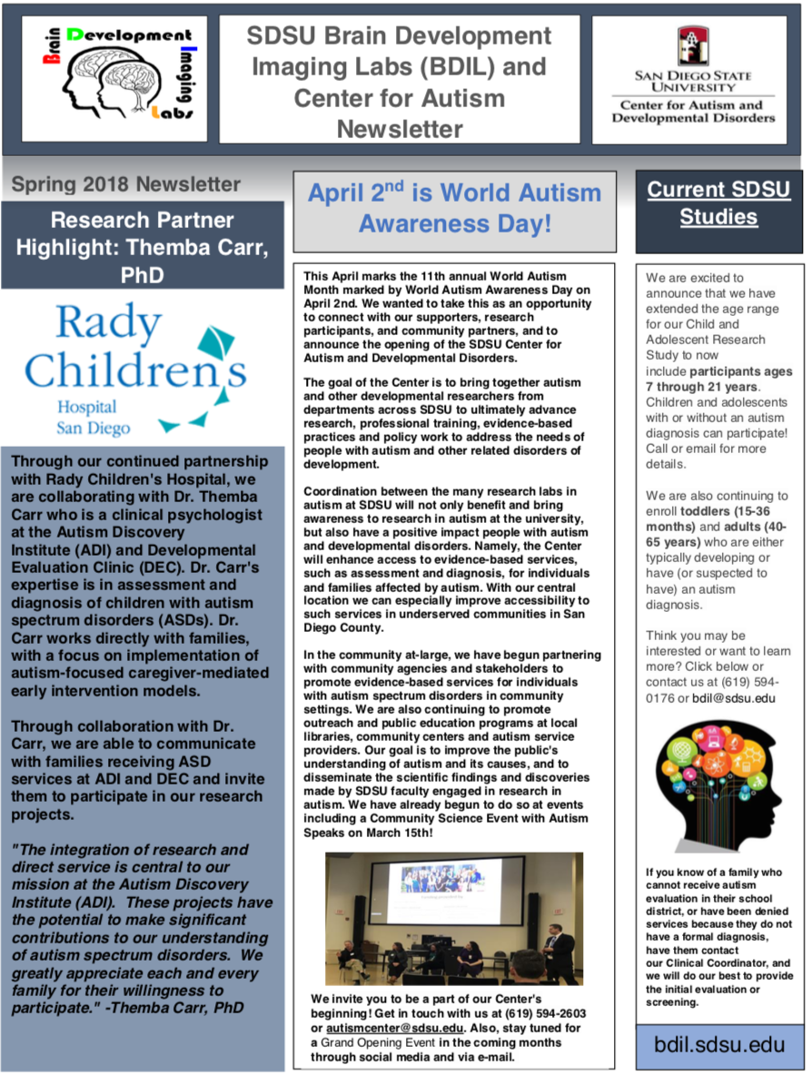
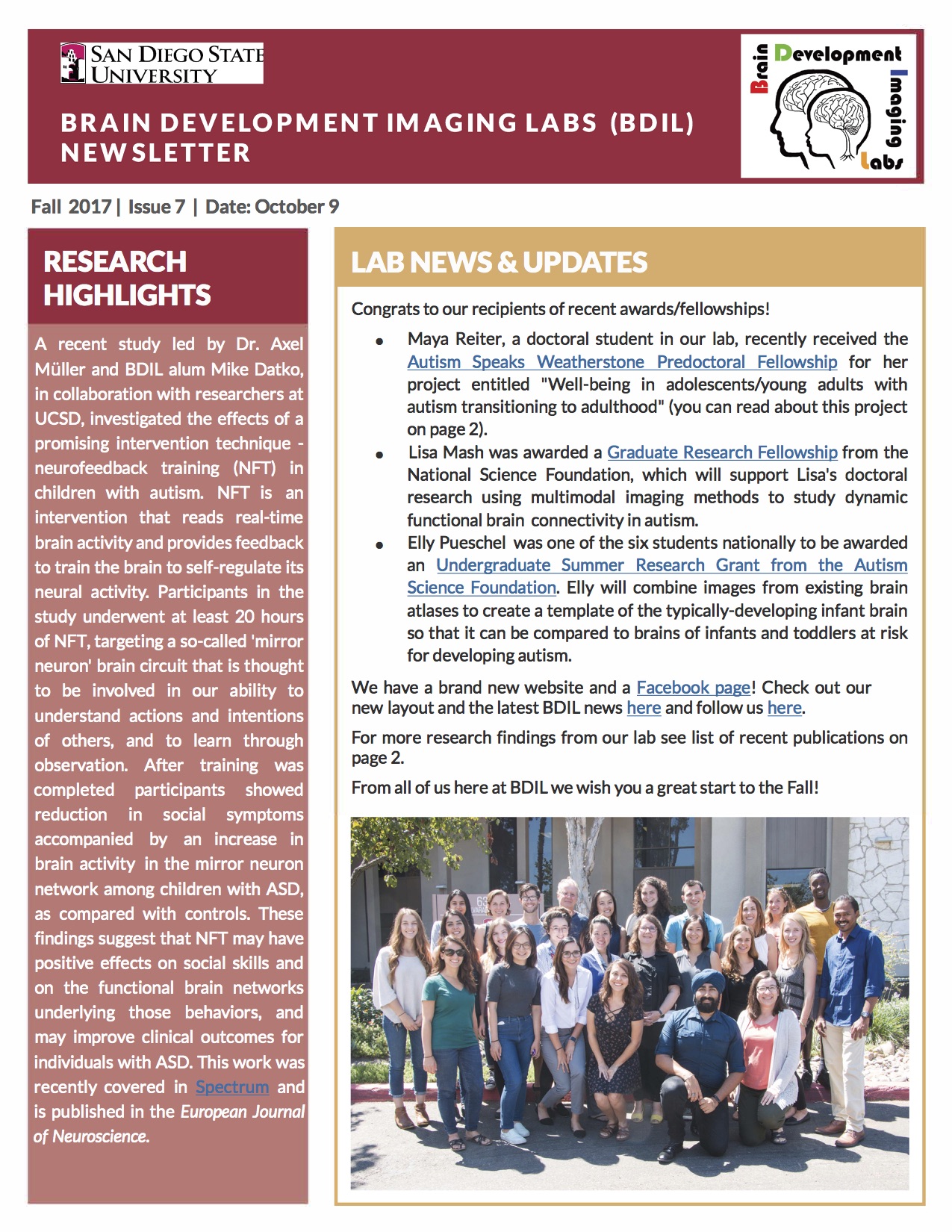
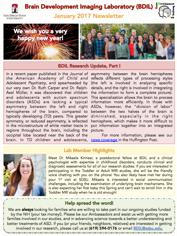
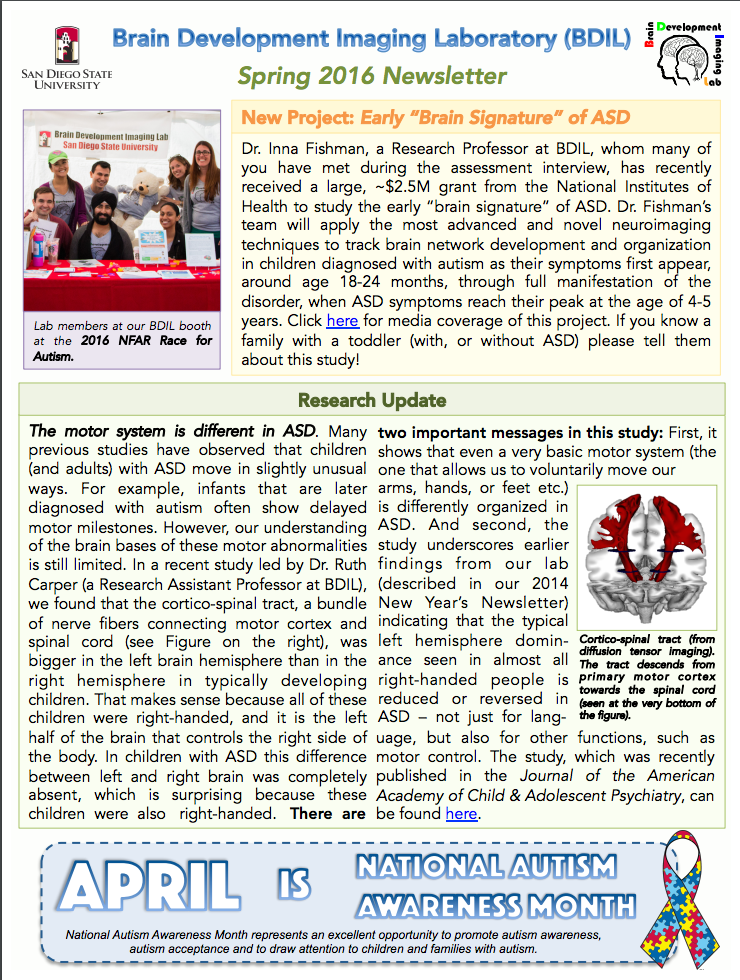
About the Brain Development Imaging Laboratories
BDIL was founded in 2001 by Dr. Ralph-Axel Müller and is now co-directed by Drs. Inna Fishman and Ruth Carper. Continuously funded by the National Institutes of Health and other major funding agencies, BDIL is dedicated to the study of brain development and its disorders, with specific focus on autism spectrum disorders (ASD). The primary research methods used by our group are measurements derived from neuropsychology and developmental science combined with different types of magnetic resonance imaging (MRI), including functional MRI (fMRI), functional connectivity MRI (fcMRI), diffusion weighted imaging (DWI), and high-resolution anatomical imaging (aMRI).
Much of our recent and current work concerns atypical organization of brain network connectivity in ASD, across the lifespan. Brain connectivity can be detected with fcMRI, which – roughly speaking – describes how well different parts of the brain “talk to each other”. Functional connectivity is supported by anatomical connectivity (bundles of axons that connect nerve cells in different brain regions). Anatomical connectivity can be studied using diffusion MRI. The brain also undergoes dramatic changes in gray and white matter during childhood and adolescence, which can be studied with anatomical MRI.
BDIL also applies additional MRI techniques, such as MR spectroscopy (which gives some insight into brain chemistry) and – in collaboration with other groups – electroencephalography (EEG) and magnetoencephalography (MEG). We also use various behavioral, neuropsychological, and diagnostic measures to characterize cognition and social and emotional functioning in children and adults with ASD and other developmental disorders, to understand links between brain and behavior.
If you are interested participating in one of our studies, please do not hesitate to contact us!


